You're about to create your best presentation ever

Free Fluoroscopy Powerpoint Template

Create your presentation by reusing a template from our community or transition your PowerPoint deck into a visually compelling Prezi presentation.

Fluoroscopy

Transcript: Fluoroscopy can be used in many examinations and procedures, such as barium X-rays, cardiac catheterization, placement of intravenous (IV) catheters , intravenous pyelogram, hysterosalpingogram, and biopsies. Fluoroscopy is an important tool for pacemaker surgery. It is also used in placement of eating tubes, without this, it would have failed. Fluoroscopy can be used as a diagnostic procedure, can also be used in conjunction with other diagnostic or therapeutic media or procedures. Fluoroscopy is used to assist doctors to see blood flow through the coronary arteries, so as to assess what causes arterial occlusion. Fluoroscopy assists the doctor in guiding the catheter into a specific location. Fluoroscopy is used in barium enemas, swallowing and meals, in defecating proctograms and enteroclysis. Fluoroscopy is also used in orthopedic surgery. Angiography of the leg, blood vessels and heart uses fluoroscopy, Cons Uses in the medical field Fluoroscopy Webdesign, C. (n.d.). Fluoroscopy. Retrieved October 09, 2017, from http://www.rasloimaging.com/features/fluoroscopy/ Fluoroscopy. (n.d.). Retrieved October 09, 2017, from http://veterinaryspecialtycare.com/diagnostic/fluoroscopy/ Fluoroscopy. (n.d.). Retrieved October 09, 2017, from http://www.cradiology.com/examinations/fluoroscopy.html G. (n.d.). Fluoroscopy GIFs - Find & Share on GIPHY. Retrieved October 09, 2017, from https://giphy.com/search/fluoroscopy Azad, A. (2013, May 13). X- ray Fluoroscopy. Retrieved October 03, 2017, from https://prezi.com/hy-0cgvcjkmf/x-ray-fluoroscopy/ Fluoroscopy. (n.d.). Retrieved October 05, 2017, from https://image.baidu.com/search/detail?ct=503316480&z=0&ipn=d&word=Fluoroscopy&step_word=&hs=0&pn=114&spn=0&di=46898544770&pi=0&rn=1&tn=baiduimagedetail&is=0%2C0&istype=0&ie=utf-8&oe=utf-8&in=&cl=2&lm=-1&st=undefined&cs=485724845%2C921078168&os=1587209073%2C1684835874&simid=3374906905%2C210215764&adpicid=0&lpn=0&ln=232&fr=&fmq=1507221665916_R&fm=&ic=undefined&s=undefined&se=&sme=&tab=0&width=undefined&height=undefined&face=undefined&ist=&jit=&cg=&bdtype=0&oriquery=&objurl=http%3A%2F%2Fwww.biomedsearch.com%2Fattachments%2F00%2F22%2F45%2F30%2F22453050%2F1532-429X-14-21-2.jpg&fromurl=ippr_z2C%24qAzdH3FAzdH3Fooo_z%26e3Bkt54j1fjw6vi_z%26e3Bv54AzdH3FgtiAzdH3FT5ow61f-6jws-pt4j-vw61t5ewfv7sw6-4w2gjptvAzdH3Fdd9cnaca_z%26e3Bip4s&gsm=3c&rpstart=0&rpnum What is a Fluoroscopy and Why Might You Need It? (n.d.). Retrieved October 03, 2017, from http://www.hopkinsmedicine.org/healthlibrary/test_procedures/orthopaedic/fluoroscopy_procedure_92,P07662 Center for Devices and Radiological Health. “Medical X-Ray Imaging - Fluoroscopy.” U S Food and Drug Administration Home Page, Center for Devices and Radiological Health, 2 Mar. 2017, www.fda.gov/radiation-emittingproducts/radiationemittingproductsandprocedures/medicalimaging/medicalx-rays/ucm115354.htm. Accessed 3 Oct. 2017. What is it - Fluoroscopy can result in high radiation doses, especially for complex interventional procedures. - Radiation- injuries to the skin and underlying tissues like “burns”, which will occur in a very short period after the exposure. - Radiation-induced cancers, which may occur in a long period (sometime later in life). - Soft tissue resolution is very poor. If you are pregnant, You need to notify your doctor before scheduling the exam. For other options, you may discuss with yout doctor You will be provided with a patient gown, and may asked to wear it. You will be provided a locker to place your personal belongings. You need to remove all jewelry and valuables, it's best to leave them at home. You must drink lots of fluid. You can drink only black coffee, plain tea, clear fruit juice or water . You cannot eat cream and milk. Low residue foods are best for you - Such as white bread, cream of wheat, meat (no fried food!), chicken, ham, and all defined liquids are allowed. You need to avoid raw fruits, raw vegetables, nuts, raisins, whole-wheat products, cold cuts, cheeses, hotdogs, and meat fats. If you are allergic or sensitive to medication, you need to notify the radiologist or technologist. How it works How a patient is prepared Pros Procedure sources A fluoroscope allows a physician to observe the internal structure and function of a patient. It is a study of moving body structures. It is a type of imaging technology which uses x-rays to provide detailed information in real time. It shows a continuous X-ray image on a monitor, It's just like an X-ray movie. X-ray beam is passed through the body part being examined. Fluoroscopy procedures are used to help diagnose disease, or to guide physicians in the treatment procedures. Fluoroscopy 's best used is to examine how things move through the patients' body. It is used for blood flow studies Fluoroscopy is used in many types of examinations and procedures to treat or diagnosis patients. For example : -Barium X-rays and enemas • It is not a painful procedure • There is valuable informationcan be find from this technology

Fluoroscopy

Transcript: Balocating, Jayson Russel C. Fluoroscopy prepared by:. Medical Imaging Diaz, Bon Fluoroscopy a type of medical imaging that shows a continuous X-ray image on a monitor, much like an X-ray movie. - During a fluoroscopy procedure, an X-ray beam is passed through the body The image is transmitted to a monitor so the movement of a body part or of an instrument or contrast agent (“X-ray dye”) through the body can be seen in detail. Esophagus - a muscular tube connecting the throat (pharynx) with the stomach about 8 inches long. Diagnoses: Stomach - a muscular organ located on the left side of the upper abdomen, it receives food from the esophagus. As food reaches the end of the esophagus, it enters the stomach Duodenum - the first part of the small intestine. It is located between the stomach and the middle part of the small intestine, or jejunum. Small Bowels - The small intestine or small bowel is the part of the gastrointestinal tract between the stomach and the large intestine, and is where most of the end absorption of food takes place Fluoroscopy is used in a wide variety of examinations and procedures to diagnose or treat patients. Some examples are: Benefits: Barium X-rays and enemas - help detect disease and abnormalities and diagnose symptoms such as pain, constipation or blood in the stool (to view the gastrointestinal tract). Catheter insertion and manipulation - to direct the movement of a catheter through blood vessels, bile ducts or the urinary system Angiograms - to visualize blood vessels and organs Risks: radiation-induced injuries to the skin and underlying tissues (“burns”), which occur shortly after the exposure, and radiation-induced cancers, which may occur some time later in life. System Operation The high-voltage generator and X-ray tube used in most fluoroscopy systems is similar in design and construction to tubes used for general radiographic applications. X-ray Source located between the X-ray tube exit port and the collimator. Added aluminum and/or copper filtration can reduce skin dose at the patient’s entrance surface Beam Filtration Shutters that limit the geometric extent of the X-ray field are present in all X-ray equipment. In fluoroscopy, the collimation may be circular or rectangular in shape, matching the shape of the image receptor. Collimation Patient tables must provide strength to support patients and are rated by the manufacturer for a particular weight limit. It is important that the table not absorb much radiation to avoid shadows, loss of signal and loss of contrast in the image. Patient Table and Pad Carbon fiber technology offers a good combination of high strength and minimal radiation absorption, making it an ideal table material. Foam pads are often placed between the patient and the table for added comfort, yet with minimal radiation absorption. Typical grid ratios range from 6:1 to 10:1 Anti-Scatter Grid an electronic device that converts the X-ray beam intensity pattern (aka, the “remnant beam”) into a visible image suitable for capture by a video camera and displayed on a video display monitor. Image Receptor — X-ray Image Intensifier (XRII) displaying images at a high frame rate, typically 25 or 30 frames per second. Image Display

Fluoroscopy

Transcript: What is Fluoroscopy? Pros: - Provides detailed internal view of the body (blood vessels, organs) - Creates a stronger understanding of an internal area - Benefits ones overall health When it comes to the overall moral and ethical generalization of fluoroscopy, it obtains a good reputation. The fluoroscopic procedure began in the nineteenth century. Since then, it has developed a strong contribution to the medical field. It has assisted physicians with the placement of devices within the body, with surgery(joint replacements, etc), helped identify direction of movements and with the overall visualization of blood vessels and organs. Fluoroscopy is definitely considered beneficial, but there is however, one aspect that raises some concern; which leads us back to radiation. In the past, the use of the fluoroscope has sparked some questions, as there is a negative stigma on the use of the radiation, to some extent of course. A considerably large population of people both in and out of the medical field question whether it is safe to participate in a fluoroscopic process, as it is believed to be hazardous to the patients and practitioners. On the other side of the equation, there is a vast amount of people who believe that the probability of being harmed by radiation is low, and withhold a strong sense of confidence with the procedure. Pros/Cons Effects of fluoroscopy on the human body Moral/Ethical Issues The primary effect that a fluoroscopic procedure can have on the human body is radiation exposure. The process of fluoroscopy is essentially the use of radiation, which can be dangerous and problematic to an individuals body if not handled correctly. Depending on the dose of radiation one may receive during fluoroscopy, there is a constant concern of being harmed by the implementation of radiation. There are two main potential effects: Radiation burns: There is a chance that the radiation flowing through your body can burn the underlying skin. Radiation induced cancers: After the fluoroscopic procedure, there is an unlikely, though possible chance of someone obtaining a cancer later in life These effects have the potential of occurring, fluoroscopy is generally a safe procedure. Cons: - Risk of harmful effects from radiation (burns, cancer) - Inflicts burden on environment Jillian Cavuoti Fluoroscopy is a type of medical procedure that provides an x-ray image of a part of a body on a monitor. It is essentially the same as taking a regular x-ray, however the result is continuous, as you are able to see the movement of the body part. The procedure is done using a fluoroscope. The machine sends out continuous or pulsing x-ray beams that take multiple shots of the organ/select area of the body, allowing it to be viewed with movement, known ultimately as an 'x-ray video'. Fluoroscopy enables physicians to assess a part of the body in greater detail. Environmental Impacts When evaluating the possible environmental impacts of fluoroscopy, there are not many outstanding points. Although there are certain things to consider that most definitely effect the environment in some aspect. The number one component of the fluoroscopic procedure is, of course, radiation. Although these waves mainly affect an individuals health directly, it also pertains to the environment. Like any process involving radiation, fluoroscopy needs to dispose of the radioactive waste. That being said, if not disposed of properly, the effects can be very negative and last a lifetime. Improper disposal is unfortunately not uncommon and is injurious to air, soil and water quality. Apart from the typical effects on the domain around us, fluoroscopy can potentially impact ones well being. Depending on the type of injection,if any, that someone can receive prior to the fluoroscopic procedure, there is a chance that someones level of energy can be altered. Fluoroscopy

Fluoroscopy

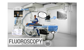
Transcript: -Fluoroscopy is used for many types of examinations -Some examples being barium X-rays,cardiac catheterization, arthrography and Biopsies Cons How it works Usage http://www.rtstudents.com/students/radiology-fluoroscopy.htm https://rpop.iaea.org/rpop/rpop/content/informationfor/healthprofessionals/1_radiology/fluoroscopy.htm https://en.wikipedia.org/wiki/Fluoroscopy http://www.fda.gov/Radiation-EmittingProducts/RadiationEmittingProductsandProcedures/MedicalImaging/MedicalX-Rays/ucm115354.htm How patient is prepared Bibliography Fluoroscopy is a technique used to get "live" X-ray images of a living patient. Radiologist uses a switch to control an X-Ray beam that is transmitted through the patient. X-rays then strike a fluorescent plate that is paired to an "image intensifier" that is coupled to a television camera. The Radiologist can then watch the images on a TV monitor. Fluoroscopy Procedure The patent is sometimes sedated for the fluoroscopic exam the patent does not have to wear a lead vest for this procedure During a fluoroscopic procedure, an X-ray beam is passed through the body. The image is transmitted to a monitor so the movement of a body part or contrast agent (“Iodine”) can be seen in detail. During a fluoroscopic procedure, an X-ray beam is passed through the body. The image is transmitted to a monitor so the movement of a body part or contrast agent (“Iodine”) can be seen in detail. In cardiac catheterization, fluoroscopy is used as an additional tool to enable the doctor to see the flow of blood through the coronary arteries in order to evaluate the presence of arterial blockages. The benefits outweigh the risks in the case of fluoroscopy Can experience blurring due to systematic lag Poses a potential for increasing the patient's risk of radiation-induced cancer Long exposure can cause radiation burns Provides real-time, moving images Display can be recorded and played back for analysis purposes Provides immediate results so patient doesn't need to wait as long for results Pros

Fluoroscopy

Transcript: Fluoroscopy Risks Fluoroscopy helped scientists better understand cellular processes because they were able to see how cellular systems operate as a whole as well as in very clear detail. Fluoroscopy allowed them to be able to see how the cells move and interact with each other and the body, which was a very big and beneficial help to the society. How does it work? By: Alia.A & Suji.U A type of medical procedure that uses x-rays to create a set of moving images (like a movie) of the patient's internal structures through a fluoroscope (type of x-ray machine that uses a continuous x-ray beam) Benefits Risks & Benefits What is Fluoroscopy? Fluoroscopy uses an x-ray beam that is passed through the body part being examined. The beam is transmitted to a TV like monitor so the body part and it's motion can be viewed in more detail. Why is Fluoroscopy used? -Lie on the table or stand -Painless procedure -You might also need to drink barium -The camera will be moved to a position above or in front of you procedure can either take a few minutes or an hour depending -Having to remove any accessory or clothing found in the area being scanned What can the patient expect? Risks radiation induced burns radiation cancer (highly unlikely) Benefits valuable + detailed information can be obtained fast and painless Morals & Ethical Issues Enhancement of the understanding of cells and cellular processes assessing stomach and bowel movement and function detecting obstructions in the airway or blood vessels It is used in many injections, treatments, examinations and procedures used to look at many body systems, including the skeletal, digestive, urinary, respiratory, and reproductive systems. used to see specific areas of the body, such as the bones, muscles, and joints, as well as solid organs, like the heart, lung, or kidneys. It's most often used to view the stomach, esophagus and upper small intestine Fluoroscopy is mainly seen as a good contribution to the medical field, because of how it helps doctors and physicians in so many ways. However some people wonder if its worth the risk to the patient even if majority of people believe that the probability of getting harmed is low. 62 ECG bpm

Now you can make any subject more engaging and memorable