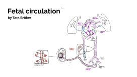
FETAL CIRCULATION
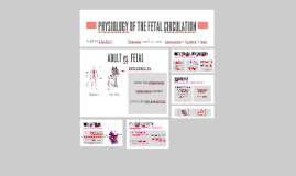
Transcript: How does the fetal circulatory system work? What if those shunts don't follow the blueprints after birth?... If you remember nothing else.... Normally closes due to increased O2 tension. Acetylcholine, bradykinins, and prostaglandin are the chemical mediators which assist in closure of the ductus arteriosus. Aortic blood is shunted into the pulmonary artery. 2-3 times more common in females than males, etiology is unknown. Most common congenital anomaly associated with maternal rubella infection during early pregnancy Premature infants often have a PDA related to hypoxia and immaturity. First line treatment is to use indomethacin to chemically close the PDA. If that is unsucessful, surgical closure of a PDA is achieved by ligation and division of the ductus arteriosus. *Adaptive fetal circulation. *Higher RBC levels. *Higher hemoglobin; up to and over 20 mg/dl. *Left-Shifted Fetal hemoglobin (HbF) p50 of 19 mm Hg compared to adult's p50 of 27 mm Hg. *Increased cardiac output (200 ml/kg/minute) FETAL CIRCULATION Josh Laack March 8, 2016 REFERENCES Patent Foramen Ovale (PFO) Patent Ductus Arteriosus (PDA) Normally closes due to increased SVR (due to separation from the placenta), increased alveolar and arterial oxygen tension with a resultant decreased PVR (due to lung expansion.) The decreased PVR leads to the decreased right heart pressures. A small, isolated patent foramen ovale essentially has no hemodynamic significance. However, if other defects are present (such as pulmonary stenosis), blood deficient in oxygen will be shunted through the foramen ovale. This hypoxemic blood will go straight into the left ventricle, producing readily identifiable cyanosis. A probe patent foramen ovale is present in up to 25% of people. Usually small enough to be considered clinically insignificant. Patent Foramen Ovale (PFO) Patent Ductus Arteriosus (PDA) PDA is considered more serious than PFO Thanks for your time, and enjoy the rest of your day! Fetal Circulatory Changes In a ....... "Cardiovascular System." Human Embryology Organogenesis. Universities of Fribourg, Lausanne, and Bern (Switzerland)., n.d. Web. 26 Feb. 2016 <http://www.embryology.ch/anglais/pcardio/umstellung02.html>. "Circulatory Changes at Birth." Fetal Circulation. University of California, Berkeley, n.d. Web. 26 Feb. 2016. <https://mcb.berkeley.edu/courses/mcb135e/fetal.html>. Brice, Glenn (2016). Advanced Human Physiology notes. University of Wisconsin La Crosse. Morgan, G.E., Mikhail, M.S. & Murray, M.J. (2013). Cliinical Anesthesiology, Fifth Editon. New York: McGraw-Hill. In the fetus, gas exchange occurs in the placenta. The fetal circulation is "shunt-dependent." The presence of fetal hemoglobin and a high combined ventricular output (CVO) help maintain oxygen delivery in the fetus despite low oxygen partial pressures. The transition from fetal to neonatal life involves closure of circulatory shunts and acute changes in pulmonary and systemic vascular resistance. Overview of Fetal Circulatory Development The system begins to develop toward the end of the third week after conception. The heart starts to beat at the beginning of the fourth week. The most critical period of heart development is from day 20 to day 50 after conception (in a time frame where many women aren't even aware they are pregnant yet...) Many crucial steps occur during cardiac and pulmonary development, and any deviation from this "normal pattern" can cause congenital heart defects, as well as pulmonary complications. 1) Ductus Arteriosus Allows blood to bypass the pulmonary circulation, connects to the descending aorta. Acts to protect the lungs against circulatory overload. Closure starts approximately 10 hours after birth, and is usually complete roughly 3 weeks later. 2) Foramen ovale Shunts highly oxygenated blood from the right atrium to the left atrium. Should close within several hours after birth. 3) Ductus Venosus The fetal blood vessel connecting the umbilical vein to the IVC Carries mostly highly oxygenated blood. Closes mechanically with cord clamping at birth. A stressed newborn can quickly revert back to fetal circulation by reopening the ductus arteriosus and foramen ovale, which can be detrimental to the neonate. Stress factors which anesthesia can help to control include: hypoxemia, hypotension, hypothermia...(3 hypos), as well as hypercapnea and hypervolemia...(2 hypers). Chronic Hypoxia - How Does the Fetus Cope? 3 Shunts in the Fetal Circulation The fetus is connected by the umbilical cord to the placenta, which is the organ that develops and implants in the mother's uterus during pregnancy. Through the blood vessels in the umbilical cord, the fetus receives all the necessary nutrition, oxygen (as fetal lungs do not pacipate in oxygen exchange), and life support from the mother through the placenta. Waste products and carbon dioxide from the fetus are sent back through the umbilical cord and placenta to the mother's circulation to be eliminated.


