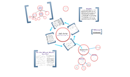
You're about to create your best presentation ever

Cell Cycle Powerpoint Template

Create your presentation by reusing a template from our community or transition your PowerPoint deck into a visually compelling Prezi presentation.

Cell Cycle

Transcript: Please note that this Prezi contains only the topics specified in class, and not all of the steps of the phases of the Cell Cycle. Thank you. Cyclins are proteins that associate with cyclin-dependent protein kinases (CDKs) to regulate their activity and the progression of the cell cycle through specific checkpoints. Cyclin D, when it interacts with CDK4/6, and Cyclin E, when it interacts with CDK2, form two complexes with very important jobs. Cyclin D is produced when growth factors stimulate Raf/MEK/ERK. A signal is sent through a MAPK signaling complex which activates c-myc, which causes the transcription of Cyclin D and other important genes. In this way, so long as growth factors are present, Cyclin D is synthesized. CDC25a, a phosphotase and CDK activating kinase (CAK), activates CDK4,6 by removing two phosphates from the CDKs when Cyclin D is expressed. (It also activates CDK2 by removing two phosphates.) Once they are both active, Cyclin D and CDK4,6 are combined to make a complex. Cyclin D is inactivated by the presence of cyclin-dependent kinase inhibitor proteins (CKIs). Cyclin D is inactivated and begins the process of degradation when one of these CKIs binds to the Cyclin D/CDK4,6 complex. Cyclin E interacts with CDK2, forming a complex that helps the cell to progress from G1 Phase to S Phase. It phosphorylates p27, an inhibitor of Cyclin D, tagging it for degradation and promoting Cyclin A, further allowing the cell to make progress towards S Phase. Cyclin E is targeted for destruction by the proteosome through ubiquitination when associated with a complex of proteins called the SCF, which is an E3 ligase that causes the ubiquitination of cyclin dependent kinase inhibitors (CKI) P27, P21, and Cyclin E. One of the most important jobs these two complexes have is regulating pRB, a tumor suppressor protein. They regulate pRB by phosphorylating it, freeing E2F. Cyclin D/CDK4,6 partially phosphorylates it, and Cyclin E/CDK2 finishes phosphorylating it. When dephosphorylated in G1, pRb binds to E2F and prevents it from doing its job. E2F, an S-phase transcription activator, starts the transcription of genes for S-phase proteins such as cyclins, cyclin dependant kinases, checkpoint regulators, DNA polymerase, and other DNA repair and replication proteins. This pushes the cell into S Phase. S Phase Cyclin A is an S-phase cyclin that insures that DNA is replicated only once. It is created in the cell after E2F is released from PRB since E2F is the transcription factor for Cyclin A. Cyclin A in turn phosphorylates E2F to deactivate it stopping its own transcription. Before the transcription of Cyclin A, Cdc6 was present in the nucleus because it acts with Pre-RC to initiate DNA replication. Cdc6 is deactivated by phosphorylation. Cdk1 and Cyclin A form a complex that phosphorylates Cdc6 hence stopping DNA replication. The Cdk1-Cyclin A complex also phosphorylates B-mib. B-mib is the transcription factor of Cyclin B. G2 Phase P53 is inactive when it is in a bond with MDM2. The MDM2-P53 protein complex is usually present in the cell and only becomes active when P53 is phosphorylated. P53 is phosphorylated when the DNA is damaged. The active P53 can either a.) go on to initiate intrinsic apoptosis (covered later) or b.) act as a transcription factor for WAF 1 and CIP 1. The resulting protein is P21. P21 binds to CDK2 and CDK1. When P21 is bound to CKD1, CDK1 cannot bind to Cyclin B and form mitosis promoting factor (MPF). MPF is necessary for the cell to proceed from the G2 phase to mitosis. CDK2 normally forms a complex with Cyclin E to phosphorylate P27, but when CDK2 is bound to P21 it cannot perform these functions. When P27 is left unphosphorylated and inactivated Cyclin D is not degraded and the cell cycle cannot proceed from the G1 phase. 1.) When the cell is infected ctls will activate the FAS ligand embedded in the membrane. The activated FAS ligand relases Caspase 8. Caspase 8 can either directly cleave Caspase 3 or it can cleave Bid. 2.) The ctl can cause perforin to punch a hole in the cell membrane and allow Grandase B to enter the cell if the mdcl is bound to a viral protein on the surface of an infected cell. Grandase B then cleaves Pre-Caspase 3 initiating apoptosis. What does Caspase-3 do? Caspase-3 cleaves proteins that then proceed to destroy the cell. One of these proteins is called ROCK 1; once a subunit of ROCK 1 is cleaved away by Caspase-3, ROCK-1 phosphorylates actins in the cell membrane causing apoptotic blebbing. Caspase-3 also cleaves ICAD away from CAD(Caspase Activated DNAase). The activated CAD travels to the nucleus and starts to chop up the DNA. M Phase/Mitosis To Start Mitosis: Cyclin B forms a protein complex with Cdk1. This complex is called MPF. MPF has two sites where it can be phosphorylated: the Y15 and T161 sites. MPF is activated when it is phosphorylated only at the T161 site. PLK1 can either cleave a phosphate from MPF directly or it can activate Cdc25 which will

Cell Template

Transcript: Nucleus A Nucleus controls all the functions of the cell for example nuclues in real life is the nucleus of human skin cells. Nuclues contains all of our genetis information and only genes that are necessary for skin cells. TIMELINE Key milestones and deliverables in the agile development timeline. Vacuole Cell walls Ribosomes Cell walls are plants, fungi, protists particularly molds and algae and most bacteria A vacuole is a membrane-bound cell organelle. In animal cells, vacuoles are generally small and help sequester waste products. In plant cells, vacuoles help maintain water balance. Ribosomes are sometimes referred to as organelles, but the use of the term organelle is often restricted to describing sub-cellular components that include a phospholipid membrane, which ribosomes, being entirely particulate, do not Lysosomes Mitochondria Mitochondria are unusual organelles. They act as the power plants of the cell, are surrounded by two membranes, and have their own genome. They also divide independently of the cell in which they reside, meaning mitochondrial replication is not coupled to cell division. membrane-enclosed organelles that contain an array of enzymes capable of breaking down all types of biological polymers—proteins, nucleic acids, carbohydrates, and lipids. Cell Template Cell membrane Agile Development A cell mambrane is like a jail cell Cell membranes serve as barriers and gatekeepers. They are semi-permeable, which means that some molecules can diffuse across the lipid bilayer but others cannot. Small hydrophobic molecules and gases like oxygen and carbon dioxide cross membranes rapidly. Nuclear Mambrane Nuclear Membrane is a double layer the encloses the cell's nucleus. The nuclear menmbrane serve to separate the chromosomes from the cell's cytoplasm and other context. Cytoskelton Strategic conclusions and future directions for agile development. Nucleolus An analogy for the nucleolus is that the nucleolus is like a building materials manufacturer. The job of the nucleolus is to build components of ribosomes for the cell which later are assembled to make the ribosome

Medical PowerPoint Template

Transcript: Medical PowerPoint Template Design Elements Color Schemes for Medical Presentations Font Selection for Readability Color schemes significantly affect audience understanding and retention. In medical presentations, using blue and green hues promotes calmness and trust, while contrasting colors can highlight key information and enhance visibility. Choosing the right font is crucial for comprehension. Sans-serif fonts like Arial or Helvetica are recommended as they are easier to read on screens. Always ensure that text is large enough to be legible from a distance. Incorporating Graphics and Images Layout and Structure Incorporating relevant graphics can enhance understanding and retention of complex ideas. Use high-quality images, charts, or diagrams that directly relate to the content to support the narrative without overcrowding the slide. A well-structured layout guides the audience’s eye and improves information flow. Utilize a grid system to maintain alignment and consistency, making sure to reserve space for visual elements. Balance text with images to avoid clutter. A Blank Canvas for Your Data Presentation Tips Best Practices for Delivery Content Organization in Medical Presentations Engaging Your Audience Practicing your presentation can lead to smoother delivery and reduced anxiety. Utilize appropriate body language, voice modulation, and eye contact to foster a connection with the audience, making your message more impactful. Audience engagement is critical for effective communication. Techniques include asking rhetorical questions, using relatable examples, and incorporating multimedia elements to maintain interest and encourage participation. Title Slides and Headings Introduction to Medical Presentations Title slides set the stage for your presentation and should include the topic, your name, and the date. Headings throughout the presentation guide the audience through the narrative and facilitate smooth transitions between topics, ensuring clarity and focus on key messages. Bullet Points vs. Paragraphs Handling Questions and Feedback Bullet points provide concise and digestible pieces of information, making it easier for the audience to follow along. In contrast, paragraphs may be necessary for complex concepts but should be used sparingly to maintain attention and avoid overwhelming the viewer. Practicing and Timing Your Presentation Using Tables and Charts Tables and charts effectively present quantitative data, making complex information more approachable. They facilitate quick understanding of trends and relationships within data, enhancing the audience’s ability to interpret clinical findings or statistical results. Rehearse your presentation multiple times to refine your delivery and timing. Understanding how long each section takes helps ensure that you cover all material without rushing or exceeding your allotted time. Encourage questions to create a dialogue with your audience. Responding thoughtfully to feedback shows respect for their input and enhances clarity for everyone involved, improving overall comprehension. Citing Sources and References Importance of Visual Aids Citing sources is crucial in maintaining credibility and allowing the audience to explore further. Proper referencing not only attributes the original work but also strengthens arguments presented in the medical content, supporting evidence-based practice. Visual aids play a crucial role in medical presentations by simplifying complex information. They help audiences grasp essential concepts quickly, improving retention and engagement through the use of charts, images, and videos. Overview of PowerPoint Features PowerPoint offers various features to enhance medical presentations, including templates specifically designed for medical content, the ability to incorporate multimedia, and options for animations that can illustrate processes or changes over time. Objectives of the Medical Template The medical PowerPoint template serves to streamline the creation of presentations by providing a standardized format. This ensures consistency in design and aids users in organizing their data effectively for clarity and impact.

The cell cycle & mitosis PowerPoint

Transcript: The first stage of cell division, before metaphase, during which the chromosomes become visible as paired chromatids and the nuclear envelope disappears. The first prophase of meiosis includes the reduction division. Work cited Cytokinesis (from the greek cyto- (cell) and kinesis (motion, movement)) is the process in which the cytoplasm of a single eukaryotic cell is divided to form two daughter cells. It usually initiates during the late stages of mitosis, and sometimes meiosis, splitting a mitotic cell in two, to ensure that chromosome number is maintained from one generation to the next. In animal cells, one notable exception to the normal process of cytokinesis is oogenesis (the creation of an ovum in the ovarian follicle of the ovary), where the ovum takes almost all the cytoplasm and organelles, leaving very little for the resulting polar bodies, which then die. My names Melina and my presentations about the cell cycle & mitosis. We are going to go through the complete cell cycle. Hope you enjoy! Prophase Interphase Cell division is the process by which a parent cell divides into two or more daughter cells.[1] Cell division usually occurs as part of a larger cell cycle. In eukaryotes, there are two distinct type of cell division: a vegetative division, whereby each daughter cell is genetically identical to the parent cell (mitosis),[2] and a reductive cell division, whereby the number of chromosomes in the daughter cells is reduced by half, to produce haploid gametes (meiosis). Metaphase google.com Telophase is from the ancient Greek. During telophase, the effects of prophase and prometaphase (the nuclear membrane and nucleolus disintegrating) are reversed. Two daughter nucleus form in each daughter cell, and phosphatases de-phosphorylate the nuclear lamins at the ends of the cell, forming nuclear envelopes around each nucleus.[1] Two theories as to how this happens are. Cytokinesis Telophase The second stage of cell division, between prophase and anaphase, during which the chromosomes become attached to the spindle fibers. Introduction! The cell cycle & mitosis Presentation.

Cell PowerPoint

Transcript: Structures formed by tubulin, the make up of microtubles Located near the nucleus Help to organize cell division and are only found in plant cells Organelles that convert the chemical energy in food into compounds that are more convenient for the cell to use Have two membranes "powerhouse" of the cell What are they? Prokaryotes VS Eukaryotes DNA Ribosomes Sack-like structure that stores materials such as water, salt, proteins, and carbohydrates Exists in both animal and plant cell but is bigger in plant cells due to their need of water and their inability to move for it There are some key organelles in both the plant and animal cell that help identify them. For example, centrioles are only found in animal cells while chloroplast and cell wall are unique to plant cells. Though no matter what, both contain cell membrane and carry DNA. Cell Wall Plant cells are more complicated than animal cells There is a major size difference between prokaryotic and eukaryotic cells Eukaryotic cells are larger and more complex. They also contain nuclei which is where their genetic information is stored Though, prokaryotic cells are smaller and have structures within themselves that are far less diverse and complicated than that of the eukaryotic cells. They do not have a nucleus and rather have free-floating genetic information Golgi Apparatus Cell PowerPoint Thin, flexible barrier that surrounds the cell It regulates what enters and leaves cell while also providing protection ad support It is composed of a lipid bilayer, studded with proteins, and most importantly, it is constantly moving in a fluid manner Organelle that modifies,sorts, and packages proteins and other materials from the ER for, either, storage in the cell or secretion outside of the cell Kind of like UPS Cytoplasm Nucleolus Network of protein filaments that help the cell to maintain its shape while also being involved in movements It consists of microfilaments and microtubules, distinct protein filaments that aid in the holding up of the cell Cytoskeleton Unique powerhouse of plant cells Captures the energy from sunlight and converts it into chemical energy in a process called photosynthesis Contain their own genetic information Strong supporting layer around the membrane of plant cells, which allows certain substances to pass through It's main function is to provide protection and support for the cell in question Made from fibers of carbohydrate and protein and are mostly made of cellulose, the base ingredient in paper Response:Yes, plant cells are more complicated than animal cells do to the fact that it has more structures which withhold the cell and identify it. For example, the cell wall is a feature that can distinguish a plant cell from an animal cell as well as chloroplast, the intricate power source of plant cells. Cell Membrane Cytoplasm is basically cell goo/gel It is an area of the cell outside of the nucleus or, in the case of a prokaryotic cell, the stuff that the DNA and the other cell organelles float in It keeps the organelles from crushing or hurting themselves, for it acts like a cushion Vacuole The carrier of genetic information specific to each cell in question It is found inside the nucleus, the control center of the entire cell, but more specifically is the make up chromatin it stands for deoxyribonucleic acid Animal Cells Cytoskeleton Centrioles Nucleus Mitochondria By: Julian Perez P.3 Inside a Prokaryote Claim: Control center of the entire cell Contains basically all of the cell's DNA and instructions to make proteins and other significant molecules Covered with a double membrane called a nuclear envelope Distributes important information to the whole cell Chloroplast Site where lipid components of the cell membrane are assembled, along with proteins and other materials that will leave the cell Like Tesla, for it uses small parts to make big things Consists of both smooth (isn't covered with ribosomes) and rough (covered with ribosomes) Prokaryotes are cells that don't have a nucleus. The greek origins of the word suggest that the cell evolved before a nuclei could develop. Instead, their DNA is not contained in a nucleus and they are generally less complicated than eukaryotes. This does not mean that they are "disabled" for they can still grow, reproduce, and respond to their environment, activities commonly associated with living things. Bacteria are examples of prokaryotes. Cell Membrane Small organelles filled with enzyme The "janitors" of the cell They remove organelles that are past their prime Lysosomes also break down lipids and carbohydrates so they they can be used by the rest of the cell Lysosome Ribosomes make/assemble proteins and are found floating in the cytoplasm They are smalle pieces of RNA and protein and are attached to the endoplasmic reticulum Small and dense region within the nucleolus Where the assembly of ribosomes begins Plant Cells Centrioles Endoplasmic Reticulum Plant cells consist of many organelles

Now you can make any subject more engaging and memorable